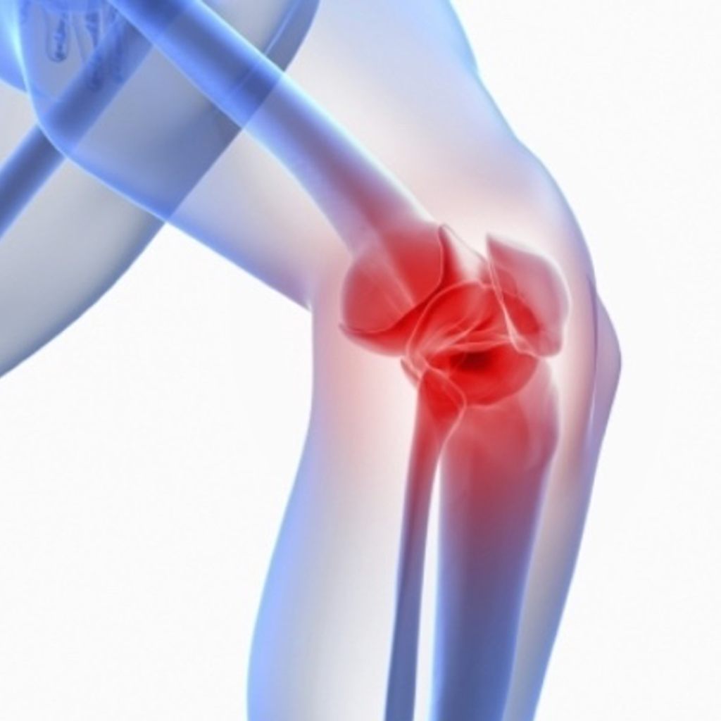
Cartilage Restoration: How surgery can help you regain movement…
The Cartilage Restoration Center is headed by Keith S. Feder, MD and Carol Frey, MD. These two orthopedic surgeons have further developed an advanced treatment center for post traumatic osteoarthritis chondral injuries and osteochondritis dissicans of the knee, ankle (talus), shoulder, elbow and hip. Dr. Feder and Dr. Frey have extensive experience in revision surgery. Procedures performed include: micro fracture, OATS (Osteochondral Autograft Transfer), ACI(Autologous Chondrocyte Implantation), osteochondral allograft implantation, and osteotomies of the femur, tibia, foot and ankle.
1. What is OATS?
Articular cartilage covers the ends of bones in joints throughout the body. Normal cartilage is smooth allowing easy gliding of the joint. When cartilage is injured, the smooth surface can become rough. On occasion, the cartilage injury exposes the underlying bone. Osteochondral grafting is a method of treating cartilage injuries that expose underlying bone. Osteochondral grafts replace both the articular cartilage on the surface and the underlying bone. The tissue can come from other parts of the patient’s body (called osteochondral autograft) or from a tissue donor (osteochondral allograft). These techniques are commonly used in the knee but can be used in other joints.
The injured area of cartilage is identified and a core of the injured cartilage and the underlying bone is removed in a method similar to coring an apple. A replacement core made up of cartilage and bone from another site in the knee (autograft) or a tissue donor knee (allograft) is then made to fit into the hole. The replacement core is gently tapped into place until it lines up with the surrounding tissue. No screws or other devices are typically needed to hold the replacement core in place since it fits tightly.
Replace both cartilage and bone with similar tissue
Patients can usually start to bear weight within 4-6 weeks of surgery. Activity is gradually increased with return to sport typically occurring after 6-9 months.
Patients often recover from both of these procedures. One advantage of these techniques is the ability to replace both cartilage and bone with similar tissue. There are limitations to the amount of tissue that can be taken from within a patient’s own knee so larger areas of cartilage loss may not be optimal for this approach. A potential concern with the use of donor tissue is the very low risk of disease transmission (like a blood transfusion). Although these techniques are too new to have data on how well patients recover in the long term, it is thought that tissue transfer has the potential to be very sustainable over time.
2. What is ACI?
Autologous chondrocyte implantation (ACI) is a relatively new, state-of-the-art procedure used to treat isolated full-thickness (down to bone) articular cartilage defects of the knee. It has been approved by the Food and Drug Administration for cartilage defects located at the end of the femur bone (thigh). ACI has also been performed for defects of the patella (knee cap) in addition to other joints of the body. Autologous chondrocyte implantation is a two-stage operative procedure.
The first procedure is performed arthroscopically in less than 30 minutes. The surgeon will harvest a small piece of articular cartilage from the patient’s knee, typically the size of one or two Tic-Tacs. This cartilage biopsy is then sent to a laboratory where the biopsy is enzymatically treated in order to isolate the chondrocytes, which are the cartilage-producing cells of the body. Once these chondrocytes are obtained, they are then expanded in number and sent back to the surgeon approximately 6 to 8 weeks later for implantation.
Physical Therapy helps with range of motion
The second-stage operation is an open procedure whereby a small patch is sewn over the articular cartilage defect. The chondrocytes that have been harvested and expanded are then injected underneath this patch where they adhere to the patient’s knee to form what is known as hyaline-like cartilage which resembles the native joint cartilage. Following implantation there is a period of restricted weight-bearing for up to 8 weeks.
During this time, physical therapy emphasizing range-of-motion of the knee and strengthening activities is prescribed. A surgeon may also recommend the use of continuous passive motion (CPM) machine to improve the graft’s success. Return to light sports activities is typically allowed at approximately 6 months with return to full sports activities between 9 and 12 months following the procedure based on the recovery. The overall success rate of ACI is approximately 85% in allowing patients to return to pain-free activities.