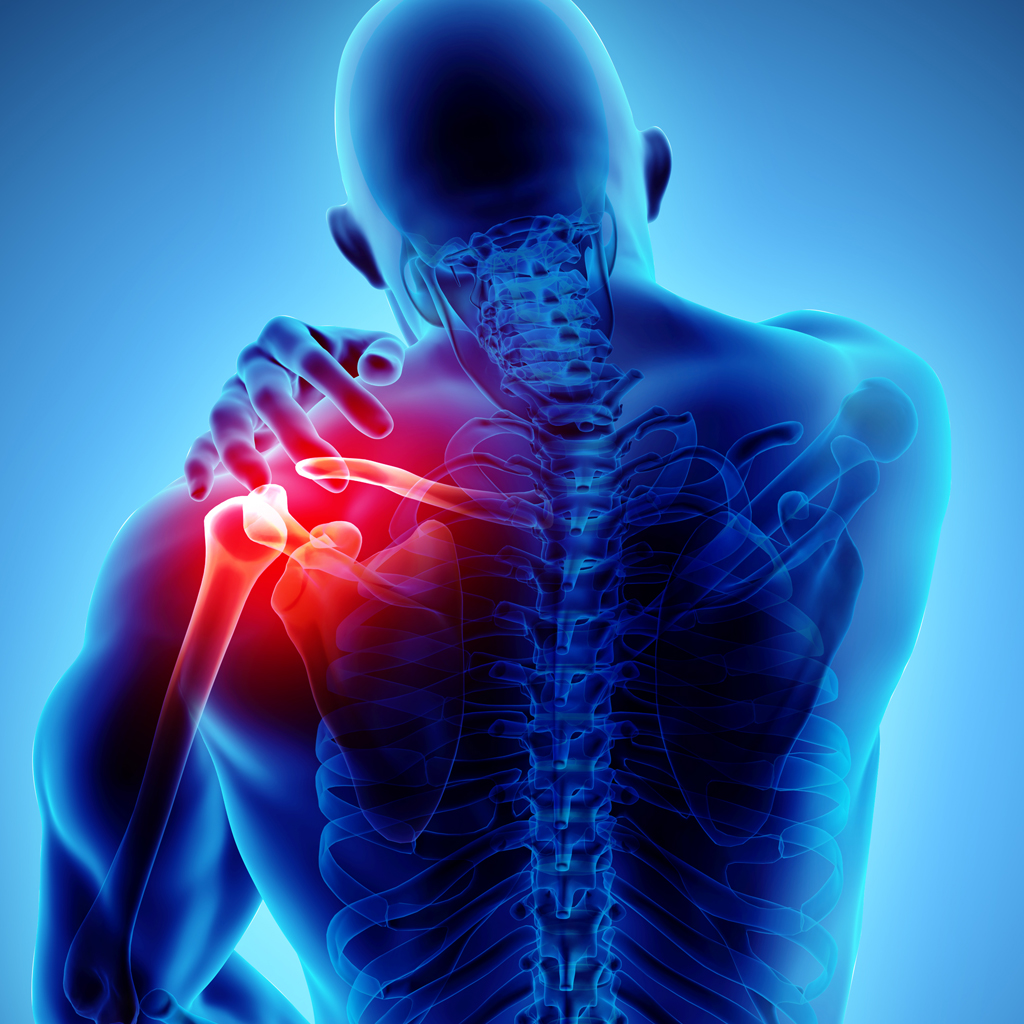
ACROMIOCLAVICULAR JOINT (AC)
AC Injuries
Acromioclavicular separations or sprains can vary in severity, depending on the extent of injury to the stabilizing ligaments and capsule. Depending on the severity of the blow causing the injury, most of the time only a partial tear of the acromioclavicular ligament will occur.
If this is the case then only a first- degree injury is produced. A second-degree injury occurs when the acromioclavicular ligament is completely tom, but the coracoclavicular ligament remains intact. This can also include subluxation or partial displacement.
The subluxation isn't always noticeable upon examination, but can be confirmed on x-ray. If the force is enough than it tears the acromioclavicular ligament, the coraclavicular ligament, and the capsule, it is known as a third-degree injury. A third degree injury is obvious on examination, and can be confirmed on x-ray.
Who
Athletes are the most likely candidates for AC injuries, more specifically football players and hockey players. These being the sports that usually put lots of stress on the shoulders in general. An athlete who has an AC injury will often leave the field/ice holding his/her arm close to the side.
It's very important to find out the exact happenings of the injury, that is did the athlete fall on the outstretched arm, or receive a severe blow to the arm (Acromial area). It is important when examining the area to rule out pain from the contusion by manipulating the clavicle at midshaft. Sometimes there's an obvious deformity or easily detected motion at the AC joint which makes it easier to diagnose the injury. The more difficult to diagnose is the less severe injury. Often times the athlete will be put through a variety of motion tests to get the proper diagnosis promptly.
Management
Treatment of these injuries depends on the severity, first and second degree sprains of the AC joint can often be treated successfully with a sling for 2 to 4 weeks, when pain is alleviated. This is usually followed up with some physical therapy to restore normal range of motion and to strengthen the upper extremities. The treatment of third-degree sprains of complete dislocations varies.
Some doctors think it best to be aggressive and perform an open reduction (surgery). Other doctors feel that they should be treated non- surgically because people tend to do well and can function with complete dislocations. When surgery is performed, it is usually directed at reconstruction of the conoid and trapezoid ligaments (coraclavicular ligaments).
Return to Activity
It is important that the athlete not return to sport until they have full range of nonpainful motion, no tenderness upon direct palpation of the acromioclavicular joint, and no pain when manual traction is applied.
Prognosis
The prognosis of these injuries is very good, obviously better for the lower grade injuries (first and second degree). However, even third degree injuries will do well if the appropriate treatment is applied and enough rehabilitation and rest is allowed.
SHOULDER PAIN
Many patients come into our office complaining of Rotator Cuff Tears, when in fact very few actually have a tear. There are several etiologies to shoulder pain; Adhesive Capsulitis (frozen shoulder), Subacromial Bursitis, Supraspinatus Tendonitis, Glenohumeral Instability, Chondrocalcinosis, Osteoarthritis, Gout, Lyme Arthritis, Bicipital Tendonitis, Ganglion Cyst are but a few possible causes of shoulder pain. It is best to think of cause of injury/pain and age in making generalizations for diagnosing shoulder pain.
Cause of Injury/Pain
Overuse
-
Subacromial bursitis
-
Supraspinatus tendonitis
-
Bicipital tendonitis
-
Impingement syndrome
Forceful Trauma
-
Glenhumeral instability
-
Adhesive capsulitis
-
Fracture
-
Rotator cuff tear
Insidious Onset
-
Lyme Arthritis
-
Adhesive capsulitis
-
Ganglion cyst
-
Tumor
Chronic
-
Gout
-
Condrocalcinosis
-
Osteoarthritis
-
Rotator cuff tear
Age
YOUNGER
Glenohumeral instability
Subacromial bursitis
Supraspinatus tendonitis
Bicipital tendonitis
Impingement syndrome
Ganglion cyst
OLDER
Lyme Arthritis
Adhesive capsulitis
Chrondrocalcinosis
Gout
Rotator cuff tear
Osteoarthritis
Diagnosing Shoulder Problems
The art of diagnosing shoulder problems is to relate the two above with a good history from the patient and a comprehensive shoulder exam. Believe it or not, there are over 50 shoulder exam tests that can be done by the examining physician alone, without the use of x-rays, MRI's, or arthrograms. The latter test may be used to confirm a clinical suspicion or in difficult cases, to aid in the diagnosis, but a patient's history of injury/pain and a good shoulder directed physical examination will usually suffice in making a diagnosis and treatment plan. Many of these problems are approached in a non-operative manner with medications, rest, ice, and physical therapy suplemented with cortisone injections on occasion. If an operation is needed, most of these conditions can be treated by arthroscopic (scope) techniques, with limited incisions and usually done as a same day surgery.
Initiating early treatment for shoulder problems is often the best curative course. If you recognize these symptoms, you should make a prompt appointment to have it evaluated further by your primary care physician or an orthopedic surgeon.
SHOULDER PAIN
Parts 1, 2, 3
SHOULDER PAIN (PART 1)
Generally, these injuries fall into three categories: Impingement Syndrome, Instability or Rotator Cuff tears. In this first article, we will discuss Impingement Syndrome. If you’ve developed shoulder pain that is worse when raising your arm, you may have this syndrome. It usually has an insidious onset, often associated with an activity that hasn’t been done for a while such as turning over garden soil, throwing a ball or household repairs. Pain with overhead activities, shoulder stiffness or a dull pain even when your not using your shoulder are the most common symptoms.
The muscles and tendons around the shoulder allow you to move your shoulder in a variety of directions including lifting, swinging, pushing or pulling and reaching. As you raise your arm overhead, a narrow space in your shoulder called the subacromial bursa gets compressed. The bursa functions normally as a fluid filled sac that allow tendons to glide without rubbing up against each other. With overuse, this bursa swells to three times its’ normal size and presses on the tendons and muscles causing inflammation and irritation. The combination of bursa swelling (bursitis) and tendon inflammation (tendonitis) is called Impingement Syndrome.
Treatment for impingement syndrome is "Active Rest". Avoid overhead activities. Ice initially to reduce inflammation followed by heat may speed your recovery. Your physician may prescribe non-steroidal anti-inflamatories. Do not stop moving your shoulder completely or a "frozen shoulder" will develop. Pendulum exercises listed below, keep your shoulder mobile without adding to the impingement.
Stir the Pot
Bend over a table and allow your affected arm to hang free. Make gentle circles with your dangling straight arm as if stirring a pot.
Bowling
In the same position as above, allow your straight arm to swing from front to back, as if throwing a bowling ball.
Saw Wood
While standing upright, grasp your affected arm with your good arm and push forward and backward as if sawing a log.
Rock the Baby
In the same position as above, grasp your affected arm and bring it across your body as if rocking a baby.
Exercises should be done for 10 minutes, 3 times a day. If the problem persists, see your doctor. Sometimes an injection of cortisone into the shoulder is needed to resolve the problem.
SHOULDER PAIN (PART 2)
In this next series, we will discuss shoulder pain related to instability. If you have dull, aching shoulder pain with the feeling that the shoulder is going to pop out of its’ socket in certain positions, you may have instability. Starting spring sports without an adequate winter training and conditioning program can lead to instability. Also excessive weight while weight-training, before the muscles are adequately built up can cause looseness of the shoulder. Finally, some people are just genetically prone to have loose joints or be "double-jointed", which is a risk factor for instability.
Instability is different from Impingement Syndrome discussed previously in that with Instability there is a provocative position which gives the sensation the shoulder is sliding out of its’ socket. This is usually associated with a sharp pain. Occasionally the entire arm my "go dead" with numbness and tingling. On occasion the shoulder actually dislocates and requires reduction either by oneself or an emergency department physician. Previous dislocations in young individuals predisposes to instability and has a high risk of further dislocation. Traditionally, gymnast, baseball pitchers, swimmers and younger athletes have loose shoulders.
Treatment for instability is aimed at conditioning and strengthening the Rotator Cuff muscles, a group of four strong muscles which encircle the upper arm and is responsible for all its movements. Formal Physical Therapy is utilized to aid in the program. A special elastic band called Theraband is used for strengthening.
Gradually strengthening is increased while maintaining the shoulder stable within its’ socket. Ice to reduce inflammation and relieve pain, followed in three days by heat to relax aching muscles and increase the blood flow (with its reparative proteins) to the shoulder is always indicated. Over the counter pain relievers such as aspirin, Tylenol, ibuprofen or naproxen can help reduce pain and inflammation.
Occasionally surgical intervention is required for repeated shoulder dislocations. These same-day surgical procedures can generally be performed arthroscopically through three tiny ½ inch incisions with special instrumentation. Surgery does allow an individual to return to his previous level of sporting activity after rehabilitation. It is best to be evaluated by an orthopedic surgeon early for this condition as recent studies have shown better success with timely intervention.
SHOULDER PAIN (PART 3)
The Rotator Cuff consists of four muscles and their associated tendons that envelop the upper arm like an upside down shoebox. These important muscles are responsible for all the movements of the upper arm and shoulder. They aid in raising your arm to comb your hair, scratching your mid-back, swinging a golf club, carrying a suitcase and throwing a ball overhand.
Rotator Cuff Injuries can result from several sources. In younger individuals, they usually are the result of a traumatic fall. In middle age individuals, they often result from repeated overuse and end-stage impingement syndrome. In the elderly, they usually result from attrition and thinning of the rotator cuff over time. Small partial tears can cause bleeding and deposits of calcium within the tendon.
Pain and weakness with shoulder use are the primary symptoms. One of the hallmarks of rotator cuff tears is pain even when lying down flat on your back. This is because without gravity the ball of the upper arm presses into the rotator cuff tear causing pain. The quality of the pain is usually sharp. Early evaluation and initiation of a treatment program is very important. Your physician will obtain a detailed history of when your shoulder hurts. He then will do a specific examination to find the location and cause of your shoulder pain. An x-ray or MRI may be ordered to evaluate your shoulder in greater detail.
Treatment initially is aimed at non-surgical treatment. The rotator cuff has the capacity to repair itself in certain situations. Resting your shoulder with a pillow between your arm and your body when sitting or lying down can help. Do not stop using your shoulder altogether as this can cause a frozen shoulder. Gentle pendulum exercises (stir the pot, bowling, rock the baby and saw wood) can keep your shoulder active while resting it at the same time. Cold and heat can sometimes help. Your physician may prescribe anti-inflammatory medications to control the pain and inflammation. A formal physical therapy program with ultrasound treatments and electrical stimulation can reduce pain levels. If the pain is severe, your physician may inject cortisone directly into the shoulder. If conservative treatment fails, surgery may be needed. Today, many rotator cuff injuries can be treated arthroscopically through three tiny incisions as same-day surgery. Talk to your doctor for more information.
SHOULDER IMPINGEMENT & TENDONITIS
Injury Description
This is one of the most commonly occurring injuries in sports where the arm is used in an overhead motion (i.e. swimming, baseball). The pain is usually felt on the tip of the shoulder or part way down the shoulder muscle. The pain is felt when the arm is lifted overhead or twisted in a certain direction. In extreme cases, pain will be present all the time and it may even wake the injured individual from a deep sleep. Throwing a baseball overhand or working overhead may become impossible.
Anatomy
The tendons of the muscles that life the arm, and the associated bursa (fluid filled sac that prevents friction) go through a very tight channel of bone (see diagram). When the arm is raised, the channel becomes smaller and makes the area very prone to inflammation. When the arm is used overhead, it can bring the asymetric bony prominence of the humerus to pinch or "impinge" against the roof of the shoulder joint. This pinches the rotator cuff and leads to tendonitis. If left too long, it can actually tear the rotator cuff.
Predisposing Factors
-
Overuse: This is the most common cause of the problem and the result of repetitive overhead motions.
-
Weak muscles: When the muscles are weak more force is exerted on the tendons and bursea causing inflammation and pain (tendonitis, bursitis).
-
Improper or inappropriate swimming or throwing techniques.
-
Strenuous training: One hard throw may start the problem.
-
Previous injuries to the shoulder.
-
Loose shoulder joint.
-
Calcium deposits.
-
Impingement of osteophytes (bone spurs) on the shoulder joint causing impingement syndrome.
Treatment
|
Rest: |
Use pain as your guide. You are only aggravating the condition if you continue your activity while experiencing pain. In very bad cases, you should refrain from using your arm for all daily activities (lifting briefcase, opening doors). |
|
Ice: |
In the early, painful stage, apply ice (frozen peas) to your shoulder twice a day for 15 minutes. Always apply ice for 15 minutes after any activity using your arm. |
|
Range: |
When use of your arm is limited, range exercises must be done twice daily. Bend at the waist and let your arm hang down. Then make large circles with your arm. These pendulum exercises will prevent your shoulder from becoming stiff. |
|
Physiotherapy: |
The physiotherapist will initially try to reduce the inflammation in your shoulder. Later, therapy will be designed to strengthen the shoulder to prevent the problem from recurring. |
|
Medication: |
Your doctor may prescribe anti-inflammation pills. These could form a very important part of the treatment. |
|
Cortisone: |
In certain circumstances an injection of cortisone into the shoulder may be indicated. It is usually a secondary treatment to supplement other therapy. After an injection you should not attempt any vigorous activities with your arm for a week to ten days. |
|
Surgery: |
Sometimes surgery is required to treat this condition. If calcium deposits occur or impingement is present, surgery may be necessary to remove these problems. |
|
Risks: |
Stiffness, infection, nerve, or blood vessel damage, bleeding, persistent symptoms, phlebitis, anesthetic problems, pneumonia, etc. MAKE SURE YOU UNDERSTAND ALL OF THESE PRIOR TO SURGERY. |
|
Rotator Cuff Tears: |
If the muscle is found to be torn at arthroscopy, an open incision may be required to repair the rotator cuff. |
Sports
General Principles
It is the overhead motion of the arm that aggravates this condition. In severe cases all sports using the arm should be avoided. When you go back to your sport, go back slowly. Throw easily and do not play for a long duration of time. Slowly increase the strength of your arm and the intensity of your game. In some sports you can avoid the overhead motions (i.e. tennis, squash).
Throwing Sports
Initially an underhand or side-arm throw will be easier than an overhand throw. Warm up well. Throw easily and gradually increase to harder throwing. Try and maintain a smooth throwing motion. This will use more of your body strength and relieve the pressure on your shoulder.
Swimming
Breast stroke or side stroke will be easier than front crawl or butterfly strokes. Sometimes the back stroke causes less pain. Perform any hard swimming (sprints) early in your swimming workout before you are fatigued. Consult a swimming coach to see if a change of your swimming style can help relieve the problem.
Exercises
Do not attempt exercising while the pain is severe. After the pain has subsided, it is of utmost importance to strengthen the shoulder muscles in order to prevent the condition from recurring. Exercise will be taught to you by your physical therapist.
SHOULDER INSTABILITY
What is it?
Shoulder instability is the abnormal relationship between the Humeral ball joint and the Glenoid socket such that there is excessive movement between the two and resultant loss of stability. This can be caused by several sources both within the shoulder joint or capsule itself, or outside the joint involving the muscles or bones. This manifest itself either subclinically by: "a feeling of looseness," or loss of momentum and strength in that shoulder.
The classic example is the baseball pitcher who loses the zing in his fastball. Other sports can include tennis serving, kayak paddle control, crew, wrestling and lacrosse. Some sports such as swimming and gymnastics actually benefit from the athletes shoulders being a little "loose."
The most obvious clinical example of shoulder instability is a dislocated shoulder. This has gone full circle from a little looseness, to stretching out the soft tissues so much that the humerus ball joint actually jumps (usually going forward) out of the glenoid socket. When shoulder dislocation occurs in a young individual (age 17-40), the is a very high probability that recurrent dislocations will occur in the future. We will talk primarily about adult instability (age 17 and up), although there is a section at the end on Pediatric Glenohumeral Instability.
Normal Anatomy
The shoulder is best thought of as a universal joint. It has a ball which is actually a cartilage sphere making up 2/3 of the top of the upper arm bone called the Humerus. It articulates with (joints) a relatively flat & oval glenoid bone, that is shaped pretty much like the racetrack at the Indy-500 – slight high riding curves at the outer edges. This flat socket is deepened by a lip of soft tissue around the entire glenoid bone called the labrum. Much like the chain-linked fence at Indy-500 deepens the racetrack to keep the cars on the track, the labrum serves to keep the ball of the humerus within the joint.
This Glenoid-labral complex functions further like the suction cup you attach to your glass window, by maintaining a negative pressure within the shoulder joint to keep their humeral ball located. Due to the flatness of the glenoid component, this makes the shoulder the most movable joint of the entire body. Freed of a matching socket for the humeral ball, such as in the hip (ball & socket joint), or a mortise to cradle the humeral head like in the ankle (hinged joint), the shoulder can achieve remarkable ranges of motion unmatched anywhere else in the body. This system makes up the static stabilizers of the shoulder joint.
The shoulder joint is actually contained with a capsule. This capsule functions like a balloon surrounding the Humeral ball and glenoid socket to keep the lubricating fluid where it needs to work. There are several regions within the capsule where it is thickened, to serve as addition restraints to the ball sliding out of the joint, dependent on the position of the arm. These ligaments are dynamic stabilizers of the shoulder joint. They move and are called to function with arm movement.
Several muscles surround the shoulder joint. Four muscles in particular come from the chest wall and back to converge on the Humeral ball. These are the rotator cuff muscles. They can be thought of as a 4-legged Tepee lying on its side. These muscles – the subscapularis, supraspinatus, infraspinatus, and terrs minor - make up the muscle stabilizers of the shoulder joint. They control a wide variety of shoulder motion including internal rotation (scratching your lower back), external rotation (opening a door), and forward flexion (reaching up). Several other important muscles make up the outer layer of shoulder stabilizers including the deltoid, pectoralis major, Latissimus dorsi, and the long head of the biceps muscle. The biceps muscle deserves special recognition, as parts of it involve all layers, and it can function as a static, dynamic, or muscle stabilizer of the shoulder depending on position of the shoulder. The anchor of the Biceps-long head is on the 12 o’clock position of the glenoid bone within the joint capsule. It then traverses over the top of the ball of the humerus where it functions to depress or hold down the ball from traveling upward and banging into the acromion or roof of the shoulder joint. As the biceps enter a small groove in the humeral head it prevents forward migration of the ball external to the shoulder joint proper. The biceps also deserves honorable mention as the usual source of shoulder pain which also radiates down the upper arm, and even sometimes involves the elbow.
Abnormal Anatomy
Shoulder instability is failure of one or more of the stabilizing systems of the shoulder. The static stabilizers can fail throughout a traumatic labral tear of either the anterior (Bankart lesion) or superior (SLAP lesion) portion of the labrum. This is usually associated with a dislocation where the arm is flung violently upward and backward (the windup phase of throwing). This can be seen when a basketball player going up for an overhead shot is stuffed by a blocker. Loss of the anterior or superior bumper allows the humeral ball to slide forward on the flat glenoid bone.
Failure of the dynamic stabilizers, namely the anterior inferior glenohumeral ligament is though to contribute to recurrent positional instability – "It bothers me only when I throw." These ligaments are probably torn or stretched at the time of the initial injury. They no longer function as a check-rein to prevent the humeral ball from sliding forward, hence recurrent instability develops. Failure of the muscle stabilizers is more complex. Causes of muscle stabilizer failure are numerous and can include inflammation (tendonitis), irritation (impingement), nerve injury due to trauma or ganglion, or rotator cuff tear.
History
A wide range of histories can be seen with instability. Usually the common denominator is a history of traumatic shoulder event that either resulted in a dislocation, or subluxation. Subluxation is the partial sliding out of the humeral ball out of the socket, such that it can easily slide back into socket with moving the arm.
Shoulder instability has been historically classified as either traumatic or atraumatic. Traumatic instability is associated with an initially normal shoulder that incurs a traumatic event that causes the shoulder to dislocate or sublux in one direction (usually anterior-inferiorly) and is almost always associated with failure of the static and dynamic stabilizers of the shoulder. There is a very high incidence of re-dislocation and recurrent instability in this group. While the first event that causes dislocation is remarkable, subsequent events are less dramatic. One patient was simply putting his arm up to place his hand behind the pillow his head was on while watching a hockey game.
Atraumatic instability is usually a systemic problem. Other joints in the body are usually loose (double jointed) as well. There may be a family history of this generalized ligamentous laxity. The patient usually has looseness in all planes of glenohumeral shoulder motion which is known as multi-directional instability of the shoulder. Sometimes these patients can make their shoulder joints pop out of place at will. Thee is usually no history of a traumatic events starting the process. There is a high degree of seeing looseness in both traumatic events starting the process. There is a high degree of seeing looseness in both shoulders. This is usually a results of the atraumatic decompensation of the muscle stabilizing group with abnormally elastic collagen within the static labrum and dynamic capsiuler ligaments. Some folks divide these groups up into simply the "torn loose" and the "born loose."
Physician Exam
Examination of the shoulder is best accomplished by exposing the entire shoulder. Wearing Tank tops assist the examiner in getting maximal benefit of the exam. It is important to assess the degree of instability. Either frank dislocation, subluxation, or apprehension can characterize recurrent instability.
Apprehension refers to the fear that the shoulder may dislocate in certain positions. This usually restricts maximal performance at a sport. The range of motion of the shoulder joint will be compared with the opposite non-involved side. Localized tenderness along the anterior glenoid rim will be sought if a labral tear is suspected. The muscles of the rotator cuff will be tested against resistance.
The apprehension test will usually be positive in patients with recurrent instability. Other special maneuvers performed by the examiner on the shoulder include the sulcus test, drawer test, push-pull test, and the fulcrum test. Finally a close assessment of the neurolgic structures will be evaluated to insure no nerve compromise.
Special Test
Many times a confirmatory test will be ordered. These include x-rays of the shoulder which is important with a history of traumatic instability. An MRI is a special machine that defines the soft tissue and bony anatomy rather precisely. Sometimes it may be necessary to add a special magnetic dye to the shoulder joint called gadolinium to view a MRI-Anthrogram. This aids in defining tears of the glenoid labrum. The drawback to MRI's is that they are performed with the arms at your side. Not in the provocative position which causes the feeling of instability. As with all special tests, they can assist in the diagnosis, but do not take the place of a well performed physical exam & history.
Differential Diagnosis
Other problems may mimic instability and are contained in the list of "other" diagnoses which may be considered, the so-called differential diagnosis list. Luckily for instability, this list is rather short and usually can be distinguished by physical exam or x-ray. Soft tissue interposition, scapular winging due to nerve palsy, seizure disorder, or electrical shock, causing violent muscle contraction with possible dislocation, tumor, and unrecognized fractures are a few causes of instability.
TREATMENT OF INSTABILITY
Conservative Treatment
This is usually the first step in restoring shoulder function. Physical therapy can assist in building up muscle and re-teaching the muscle the proper sequence to contract to restore coordinated, strong muscle contractions that are important for maintaining glenohumeral stability.
The rotator cuff muscles play an important role in stabilizing the shoulder joint and optimal control of neuromuscular forces is required to restore shoulder function. Your physical therapist will assist you in learning how to isolate individual muscles of the rotator cuff and strengthen them. This strengthening is initially performed within the "stable range" of shoulder function.
In atraumatic instability, studies have shown an 80% success rate with physical therapy. Unfortunately on 16% of patients with traumatic instability improved. Physical therapy is important even in traumatic instability to improve the muscle and tissue tone prior to planned surgery. Finally the old adage of "If it hurts doing that, don't do it!" is true here. It is important to avoid activities that stress the capsular and muscular structures. Certain habits must be broken to avoid the "unstable" positions. Any position, action, or sport that promotes shoulder subluxation or dislocation must be avoided.
Surgery
Tremendous gains have been made in the past ten years for surgery on shoulder instability. Most procedures can be performed through the arthroscope (scope) as "in & out, same-day" surgery with the use of three 1/2 inch small incisions. Bone anchors with attached suture 9thread) allow the reattachment of torn tissues. These are made in absorbable or non-absorbable materials. My preference is to use absorbable suture anchors in younger athletes with simple tear patterns. It takes about 6 weeks for the tissues to heal to bone, so the anchors are around for plenty of time to allow for healing. The body reabsorbs the absorbable tack or suture anchor over 3 months, leaving no trace behind! Lasers or even more improved thermal controlled radiofrequency devices are used to shrink the redundant capsular tissues and stretched capsular ligaments.
In large studies performed at the U.S. Coast Guard Academy, in addition to West Point and the Navy Academy, success rates from these completely arthroscopic procedures approached the rates of older open surgical techniques at about 80% to 97% when defined by redislocation or recurrent instability after surgery. The morbidity as defined by hospitals stay, patient pain levels and return to sport was far superior in the arthroscopic group. Open techniques are still utilized for revisions, or complicated cases.
Rehabilitation
After surgery it is very important to get into a regularly scheduled physical therapy rehabilitation program. Usually the arm will be in a sling post-op with a special formfitting ice pack in foam on the shoulder. ice can be discontinued after 2 or 3 days when comfortable. Studies have shown a 50% reduction in narcotic pain medications post-operatively with the use of ice therapy. Simple pendulum exercises can be performed at home three times a day for the first two weeks. Then, depending on the repair, when you return to have the single stitches closing each wound removed you'll be enrolled in a formal P.T. programs.
The shoulder is kept "protected" for 6 weeks while soft tissue healing occurs. Then active range of motion and strengthening are begun. The goal is to have the patient return to activities of daily living by 12 weeks post-op. A more conservative 14-18 weeks is used to return the athlete back to his sport.
Frequently Asked Questions
Does it hurt?
The pain is substantially less with the arthroscopic techniques today then with older open techniques. Patients usually say it hurts for 2-3 days then relents to a dull tooth-ache like pain for 3-6 weeks. As healing occurs the pain is intermittent and often associated with the physical therapy sessions. You'll go home with strong and mild painkillers to assist you in dealing with the discomfort. Cryotherapy or the use of ice sleeves has substantially reduced the amount of pain perceived.
Will I be able to return to my sport?
The aim for these advanced arthroscopic techniques in sports medicine is to return athletes to their previous level of functioning in as rapidly amount of time that is safe for the individual. There have been football players, baseball pitchers, wrestlers, crew team, lacrosse players, swimmers, and basketball players that have returned to their sport at NCAA division-III levels. Over 90 Coast Guard Cadets have been commissioned into the U.S. Coast Guard after undergoing shoulder stabilization procedures.
I have one dislocation, and now my shoulder is just a little loose...should I be worried?
The answer to this one varies with each individual. A comparison to the opposite non-involved extremity will usually exhibit significant more shoulder laxity then was appreciated. An early evaluation by an Orthopedic Surgeon can help advise you on the proper course of treatment prior to a re-dislocation occurring.
I think I've got instability. What do I do now?
In these days of Managed Care and Health plans, most insurance plans demand that you get referred to an Orthopedic Surgeon through your Primary Care Physician, tell him your symptoms, and request a referral to an orthopedic surgeon.
SKELETAL MUSCLE CRAMPS DURING EXERCISE
The most commonly seen ailment at marathons and triathlons is skeletal muscle cramps. The causes and treatment of this physiological phenomenon has not been completely defined. Many conditions can elicit muscle cramps, such as congenital abnormalities or neuromuscular diseases, but in athletes the condition is usually exercise-associated. Establishing a thorough medical history is the most effective way to determine if the cramps are exercise related or the result of another medical condition.
Muscle cramping due to exercise is a spontaneous, painful, and involuntary contraction during the course of, or at the cessation of exercise. Exercise-associated muscle cramping does not occur in resting skeletal muscle.
The concern with muscle cramping first appeared in the early 1900’s with reports of persons cramping in hot and humid conditions, such as coal and steamship workers. This led to the current thought that bodily hydration, or lack of, and the environment play a key role in the occurrence in muscle cramping.
Recent theories suggest that exercise-associated muscle cramping is due to a neurological short circuit. It is posed that the "abnormality of sustained alpha motor neuron activity, which stems from aberrant control at the spinal level." Tired muscles cause the muscle spindle to be continually stimulated therefore causing the inhibition of the Golgi tendon organ to shut down the activity.
The muscles must commonly affected are ones that cross two joints. During activity two- joint muscles, such as the calf and hamstring, are in a shortened position during contraction. "Contraction in this state produces decreased tension in the tendons of the muscles as well as decreased Golgi-tendon activity."
The best way to decrease muscle spindle excitation is passive stretching. It helps to eliminate to cramp and increase Golgi-tendon activity thereby inhibiting the constant excitatory response. This treatment follows the theory of abnormal spinal reflex activity.
This indicates that stretching is an important factor in eliminating exercise-associated muscle cramps and that endurance events such as running can quickly tire muscles, which may lead to cramping.
Exercise-related cramps often present themselves with significant discomfort and a muscle belly that is hard to the touch from continuous contraction. Athletes do not usually show any other signs of physiologic distress, either dehydration or hyperthermia.
The most effective treatment for exercise-associated muscle cramps is stretching of the pertinent muscle or muscle group. It is best to hold the stretch until the cramp subsides and is released. If the cramps continue or are extremely severe then further medical treatment should be sought. It should be explained to the athlete that if they experience any other symptoms such as lack of urination or unusually colored urine they must get medical attention immediately.
The best way to decrease the risk for muscle cramping during intense exercise is good conditioning, proper stretching, and proper nutrition and fluid intake. The best offense is a good defense.


